If you have acute symptoms right now — pus around an implant, marked swelling, fever, or an implant that has become loose — contact your dental office today rather than waiting for your next scheduled visit. Peri-implantitis is slow in most cases, but an acute infection is not.

An implant cannot get a cavity, which is why many people assume it cannot get sick. It can. The tissue and bone that hold it in place are living structures, and they respond to bacterial biofilm much the way they do around a natural tooth. Peri-implantitis is the term for that response once it has reached the bone — and because it is usually painless, it is often found late.

What is peri-implantitis?

Peri-implantitis is an inflammatory condition affecting the soft tissue and the supporting bone around an osseointegrated dental implant. It is defined by three findings taken together: bleeding or suppuration when the tissue is gently probed, increased probing depth compared with earlier measurements, and radiographic bone loss beyond the small amount of remodeling that normally occurs in the first year after placement.

Large meta-analyses put the prevalence at roughly 22 percent of implants, with the milder precursor condition affecting around 43 percent. Those numbers are high enough that anyone with implants should treat maintenance as part of the treatment rather than as an optional add-on.

Peri-implant mucositis versus peri-implantitis

This distinction determines everything that follows, because one condition is reversible and the other is not.

Peri-implant mucositisPeri-implantitis
Tissue involvedSoft tissue onlySoft tissue and bone
Bleeding on probingYesYes, often with pus
Bone loss on X-rayNone beyond initial remodelingProgressive
Approximate prevalence~43% of implants~22% of implants
ReversibleYes, with professional cleaning and improved home careNo — bone that is lost does not fully return
Analogy in natural teethGingivitisPeriodontitis

Mucositis is the window in which the problem can be solved completely. That is the practical reason for recall appointments: not to clean an implant that looks fine, but to catch the stage that still responds to cleaning.

Does peri-implantitis hurt?

Usually not, and this is the most dangerous thing about it. Unlike a natural tooth, an implant has no periodontal ligament — the fiber suspension that carries nerve endings and reports pressure and inflammation. Without it, an implant can lose a substantial amount of surrounding bone while feeling entirely normal to bite on.

When pain does appear, it typically means one of three things: the inflammation has become acute with pus formation, the surrounding soft tissue is swollen and pressing against the crown or the abutment, or bone loss has progressed far enough that the implant has begun to loosen. Discomfort with a dental implant is therefore never something to observe and see whether it settles. Mild tenderness when brushing, a dull pressure, or gums that feel sore in one specific spot all warrant an appointment.

Symptoms: what to watch for

  • Gums around the implant that bleed when brushing or flossing
  • Redness, puffiness or a bluish-purple tinge in the tissue at the implant
  • Pus at the gum margin, or a bad taste that keeps coming back
  • Persistent bad breath localized to one area
  • Gum recession, making the implant collar or the metal thread visible
  • A crown that suddenly looks longer than it used to
  • Food packing into a space that did not exist before
  • Tenderness or pressure when chewing on that side
  • Any perceptible movement of the implant — this is a late sign

The last point deserves emphasis. A natural tooth loosens gradually as bone is lost. An osseointegrated implant stays rigid until the very end and then becomes mobile abruptly. Mobility means the implant has lost integration and it will not re-integrate.

What does peri-implantitis look like?

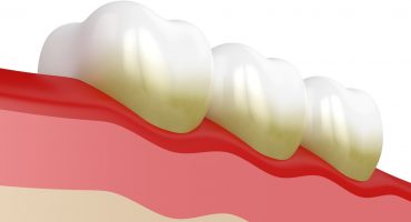
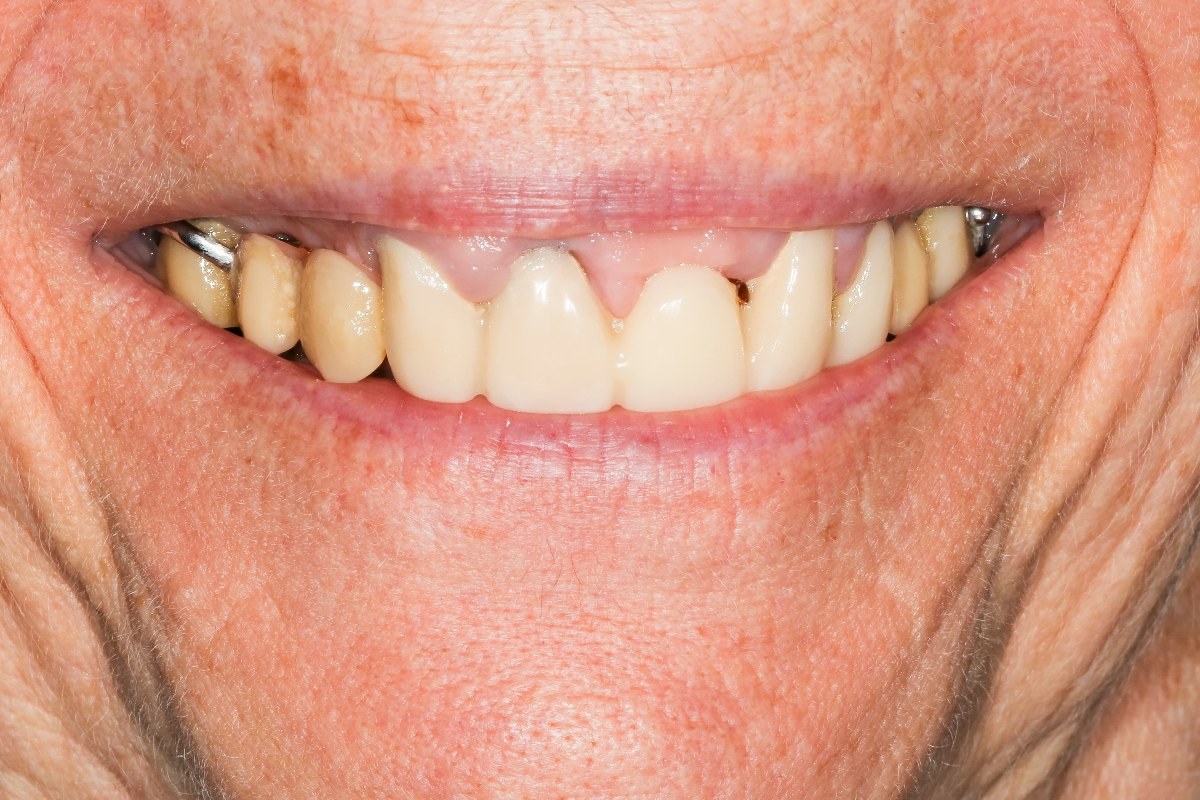
In the mouth, the earliest visible change is color and texture: healthy peri-implant tissue is pale pink and firm, inflamed tissue is red or dusky, glossy and swollen, and it bleeds readily. As the condition advances, the gum margin migrates downward and the polished implant neck — or, in more advanced cases, the rough threaded surface — becomes visible. Pressing gently along the gum may express a bead of pus.

On an X-ray the picture is more definitive: a crater-shaped or saucer-shaped radiolucency around the implant, widest at the top and tapering downward, in contrast with the straight, even bone level seen around a healthy implant. Your dentist compares this against the baseline film taken when the implant was placed, which is why that first radiograph matters years later.

Stages of peri-implantitis

There is no single universally adopted staging system, but clinicians commonly describe progression in terms of how much of the implant has lost bone support:

Early. Bleeding on probing, probing depths increased to roughly 4 to 5 millimeters, bone loss under 25 percent of the implant length. Usually no symptoms at all.

Moderate. Probing depths of 6 millimeters or more, suppuration frequently present, bone loss between 25 and 50 percent. Recession and visible implant threads are common. Still often painless.

Advanced. Bone loss exceeding 50 percent, deep pockets, persistent pus, and eventually mobility. At this point implant removal is usually the realistic option.

Because the early and moderate stages are silent, the stage at diagnosis is largely determined by how regularly the implant is checked, not by how bad the biology is.

What causes peri-implantitis?

The primary cause is bacterial biofilm accumulating at the implant–tissue junction. Everything else on this list changes how easily that biofilm forms and how strongly the body reacts to it.

  • A history of periodontitis. The strongest single predictor. Patients who lost their natural teeth to gum disease carry both the bacterial profile and the host response that caused it.
  • Inadequate plaque control, often because the restoration is shaped in a way that makes cleaning difficult.
  • Smoking, which impairs blood flow to the peri-implant tissue and blunts healing.
  • No maintenance program. Implants placed and then never professionally reviewed show markedly higher rates.
  • Residual cement from a cemented crown, left below the gum line, where it acts as a permanent plaque trap. This is a common and entirely preventable cause, and it is the main argument for screw-retained restorations.
  • Poor implant position or prosthetic design, including over-contoured crowns and bridges that cannot be cleaned underneath.
  • Uncontrolled diabetes and other conditions affecting immune response and wound healing.
  • Lack of keratinized tissue around the implant, which makes brushing uncomfortable and therefore less thorough.
  • Occlusal overload, particularly in patients who grind, which is a contributing rather than an initiating factor.

Is peri-implantitis reversible?

Partly, and the honest answer depends on what stage you are asking about. Peri-implant mucositis is fully reversible: remove the biofilm, improve home care, and the tissue returns to health.

Peri-implantitis is not reversible in the sense of restoring what was lost. Bone that has resorbed does not spontaneously regrow. What treatment can achieve is arrest — stopping the progression, eliminating bleeding and suppuration, and reducing pocket depth so the site becomes maintainable. In favorable contained defects, regenerative surgery can rebuild a portion of the lost bone, but complete restitution to the original level is not a realistic expectation and should not be promised.

The practical goal is a stable implant that can be kept clean for many more years. That is achievable in a substantial proportion of cases treated at the early and moderate stages.

How peri-implantitis is treated

Non-surgical therapy

The first step is always mechanical disruption of the biofilm. This is done with instruments that do not scratch the implant surface — titanium or PEEK-tipped curettes, air-polishing devices using glycine or erythritol powder, and ultrasonic tips with non-metallic coverings — combined with antiseptic irrigation, usually chlorhexidine. If the crown is removable, taking it off gives far better access and is worth doing.

At the same time the cause is addressed: residual cement is removed, an over-contoured crown may be reshaped or remade, home care technique is retrained with interdental brushes sized to the site, and smoking cessation is discussed. Non-surgical therapy alone reliably resolves mucositis. For established peri-implantitis it improves the situation but often does not fully resolve deep defects, which is why the site is reassessed after six to eight weeks.

Surgical therapy

If bleeding and deep pockets persist, surgery gives direct access to the implant surface. Two broad approaches exist. Resective surgery reduces pocket depth by recontouring the bone and repositioning the gum, and may include smoothing the exposed implant threads; it is effective and predictable but leaves the implant visibly longer, which matters in the front of the mouth. Regenerative surgery fills the defect with a bone substitute and often a membrane, aiming to rebuild support; it works best in deep, contained, circumferential defects.

Both depend on thorough decontamination of the exposed implant surface, which is technically demanding because the roughened surface designed to hold bone also holds bacteria.

Antibiotics

Antibiotics are an adjunct, never a stand-alone treatment. Systemic antibiotics are reserved for acute infection with swelling, pus and systemic signs, or for medically compromised patients. Locally delivered antimicrobials placed directly into the pocket are sometimes used alongside mechanical debridement. Prescribing antibiotics without removing the biofilm produces a temporary improvement and a return of symptoms after the course ends.

When the implant has to be removed

Removal becomes the right decision when the implant is mobile, when bone loss exceeds roughly half the implant length, when infection recurs despite properly executed treatment, or when the implant sits in a position that cannot be cleaned or restored acceptably. Explantation is usually straightforward, and after healing — often with bone grafting — a new implant can frequently be placed. It is worth saying plainly: replacing a failing implant early, in a controlled way, generally produces a better long-term result than defending one that has already lost most of its support.

Do ceramic implants get peri-implantitis?

They can. Zirconia is not immune to bacterial colonization, and the biological mechanism is the same. There is laboratory and early clinical evidence that zirconia surfaces accumulate less biofilm than titanium and that the surrounding soft tissue may respond favorably, but the long-term comparative data are still limited and the difference should not be presented as protection. For a fuller comparison of the two materials, see the articles on ceramic implants and titanium implants.

Prevention: what actually works

Maintenance visits every three to six months, with probing and periodic radiographs, are the single most effective measure — they catch mucositis while it is still curable. Daily cleaning of the implant–gum junction with an interdental brush or a water flosser matters more than the toothbrush itself. Screw-retained crowns avoid the cement problem entirely. Smoking cessation produces a measurable reduction in risk. And if you lost your natural teeth to periodontitis, that condition must be treated and stable before implants are placed, and monitored more closely afterward.

Frequently asked questions

What is peri-implantitis?

It is inflammation around a dental implant that has progressed into the supporting bone, causing bleeding, deepening pockets and progressive bone loss. It is the implant equivalent of periodontitis and affects roughly one in five implants.

Does peri-implantitis hurt?

Usually not. Implants lack the nerve-carrying ligament that natural teeth have, so significant bone loss can occur without discomfort. Pain generally indicates an acute infection or an advanced stage, which is why it should always be assessed promptly.

What are the first signs of peri-implantitis?

Bleeding when brushing around the implant, redness or swelling of the gum, and bad breath or a bad taste in one specific area. Visible thread or a crown that appears to have lengthened are later signs.

Is peri-implantitis reversible?

The earlier stage, peri-implant mucositis, is fully reversible. Established peri-implantitis can be arrested and the site made stable and cleanable, but lost bone does not fully regrow. Regenerative surgery can rebuild part of it in favorable defects.

How fast does peri-implantitis progress?

Progression is generally faster and less linear than in natural teeth, with periods of relative stability interrupted by episodes of rapid loss. This unpredictability is the reason for regular monitoring rather than symptom-driven visits.

Can peri-implantitis be treated without surgery?

Mucositis, yes. Established peri-implantitis often needs surgical access to decontaminate the implant surface properly, especially where pockets exceed 6 millimeters. Non-surgical therapy is always the first step and is reassessed after six to eight weeks.

Will my implant fall out?

Not without warning signs that a dentist can detect long before it happens — which is the argument for regular checks. An implant that has become mobile has lost osseointegration and needs to be removed; it will not re-attach.

Can I get a new implant after one fails?

In most cases yes. The site is allowed to heal, bone is grafted if needed, and a new implant is placed. Success rates for replacement implants are good, provided the original cause — biofilm control, smoking, untreated gum disease, a cleanable restoration design — has been addressed.

Medically reviewed: This article was reviewed for medical accuracy by a dentist within the Leading Implant Centers network.

Sources

Certified implantologists from our network

Every practice in our network is admitted only after meeting the quality criteria of Leading Implant Centers.

Find an implantologist near you

Our Scientific & Advisory Board developed these admission criteria and takes part in reviewing new member practices. Quality criteria · Scientific & Advisory Board